


CT scans may increase brain cancer risk


- Created on Saturday, 30 June 2012 00:00
- Last Updated on 18.02.2013
- Published Date
Children and young adults scanned multiple times by computed tomography (CT), a commonly used diagnostic tool, have a small increased risk of leukemia and brain tumors in the decade following their first scan. These findings are from a study of more than 175,000 children and young adults that was led by researchers at the National Cancer Institute (NCI), part of the National Institutes of Health, and at the Institute of Health and Society, Newcastle University, England.
Researchers from the National Cancer Institute and Institute of Health and Society at Newcastle University in England reviewed 175,000 cases and found that in children ages ten and younger, two to three scans can triple the risk of brain cancer and five to ten scans can triple the risk of leukemia.
The researchers emphasize that when a child suffers a major head injury or develops a life-threatening illness, the benefits of clinically appropriate CT scans should outweigh future cancer risks. The results of the study were published online in The Lancet on June 7, 2012.

Risk is small
Despite the elevation in cancer risk, these two malignancies are relatively rare and the actual number of additional cases caused by radiation exposure from CT scans is small. The most recent (2009) U.S. annual cancer incidence rates for children from birth through age 21 for leukemia and brain cancers are 4.3 per 100,000 and 2.9 per 100,000, respectively. The investigators estimate that for every 10,000 head CT scans performed on children 10 years of age or younger, one case of leukemia and one brain tumor would occur in the decade following the first CT beyond what would have been expected had no CT scans been performed.
CT scans deliver a dose of ionizing radiation to the body part being scanned and to nearby tissues. Even at relatively low doses, ionizing radiation can break the chemical bonds in DNA, causing damage to genes that may increase a person’s risk of developing cancer. Children typically face a higher risk of cancer from ionizing radiation exposure than do adults exposed to similar doses.
The investigators obtained CT examination records from radiology departments in hospitals across Britain and linked them to data on cancer diagnoses and deaths. The study included people who underwent CT scans at British National Health Service hospitals from birth to 22 years of age between 1985 and 2002. Information on cancer incidence and mortality from 1985 through 2008 was obtained from the National Health Service Central Registry, a national database of cancer registrations, deaths and emigrations.
Clear radiation/cancer relationship
Approximately sixty percent of the CT scans were of the head, with similar proportions in males and females. The investigators estimated cumulative doses from the CT scans received by each patient, and assessed the subsequent cancer risk for an average of 10 years after the first CT. The researchers found a clear relationship between the increase in cancer risk and increasing cumulative dose of radiation. A three-fold increase in the risk of brain tumors appeared following a cumulative absorbed dose to the head of 50 to 60 milligray (abbreviated mGy, which is a unit of estimated absorbed dose of ionizing radiation). Similarly, a three-fold increase in the risk of leukemia appeared after the same dose to bone marrow (the part of the body responsible for generating blood cells). The comparison group consisted of individuals who had cumulative doses of less than 5 mGy to the relevant regions of the body.
The absorbed dose from a CT scan depends on factors including age at exposure, sex, examination type, and year of scan. Broadly speaking, two or three CT scans of the head using current scanner settings would be required to yield a dose of 50 to 60 mGy to the brain. The same dose to bone marrow would be produced by five to 10 head CT scans, using current scanner settings for children under age 15.
Lower dosages called for
In countries like the United States and Britain, the use of CT scans in children and adults has increased rapidly since their introduction 30 years ago. Due to efforts by medical societies, government regulators, and CT manufacturers, scans performed on young children in 2012 can have 50 percent lower radiation doses, compared to scans carried out in the 1980s and 1990s, say the investigators. However, the amount of radiation delivered during a single CT scan can still vary greatly and is often up to 10 times higher than that delivered in a conventional X-ray procedure.
The lead author of the study was Mark S. Pearce, Ph.D., Institute of Health and Society, Newcastle University. “CT can be highly beneficial for early diagnosis, for clinical decision-making, and for saving lives. However, greater efforts should be made to ensure clinical justification and to keep doses as low as reasonably achievable,” said Pearce.
This study was supported by contract NO2-CP-75501 from the U.S. National Cancer Institute.
# # #
Reference: Pearce M S, et al. Radiation exposure from CT scans in childhood and subsequent risk of leukaemia and brain tumours: a retrospective cohort study. The Lancet. June 7, 2012.
Photo courtesy National Cancer Institute
Questions/comments? contact Jean Rickerson at This email address is being protected from spambots. You need JavaScript enabled to view it.
Brain Health

It is rare for a sports-related concussions to result in a more serious injury such as a skull fracture or hematoma. Nonetheless, it pays to be aware that catastrpohic injuries do sometimes occur as ...
read more...-
CT scans may increase brain cancer risk
Children and young adults scanned multiple times by computed tomography (CT), a commonly used dia...
-
Sub-concussive impacts may affect learning
MINNEAPOLIS – A new study suggests that head impacts experienced during contact sports such as ...
-
Physical activity boosts learning
INDIANAPOLIS – School administrators looking to restructure the academic schedule should consid...


Neuroscience

Athens, Ga.- University of Georgia researchers have developed a map of the human brain that shows great promise as a new guide to the inner workings of the body's most complex and critical organ.
...
read more...-
Does CTE infect neuron to neuron?
NFL Hall of Famer "Iron Mike" Webster's life ended in 2002 when he suffered a heart attack at age...
-
Progesterone seems to protect neurons after injury
It is not yet known why girls suffer concussions at a higher rate than boys. The most prevalent...
-
Amino acids may restore concussion's chemical imbalance
Concussions are often called the "invisible" injury because they are usually not detectable by t...
Resources
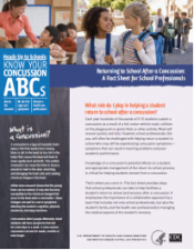
- School professionals play an important role in the health of all students. Recognizing the signs and symptoms of concussion is important, as is managing their return to school ...
- https://
- CDC's Concussion Training for Clinicians
-
Concussion Education Video Programs - Free and Easy
Parents, athletes, coaches and medical professionals have access to concussion education created...
-
New concussion guidelines for team physicians
INDIANAPOLIS – Team physicians who assess and treat athletes suspected of concussion have new ...


Help Us Help
https://https://
 Its all about the kids!
|
 |
 |
 |
 |
quick links

iBaselineTM a division of ...

Knowing the signs and ...

Every school district and ...

Implement a concussion ...

Concussion – The Basics ...

Signs and symptoms of ...

What to do if a concussion is ...

Most concussions heal within ...
What to do if a concussion is ...

...

School medical ...

American Academy of Neurology ...
Latest News
Concussions Occur...
...in Any Sport
REMOVE athlete from play
REFER to medical provider
REST no sports, no texting/TV
RETURN only with doctor's OK
Source: Children's Hospital Boston, Sports Concussion Clinic