


Brain injury's 911 call


- Created on Monday, 28 May 2012 16:32
- Last Updated on 07.12.2012
- Published Date
Like emergency workers rushing to a disaster scene, cells called microglia speed to places where the brain has been injured, to contain the damage by ‘eating up’ any cellular debris and dead or dying neurons. Scientists at the European Molecular Biology Laboratory (EMBL) in Heidelberg, Germany, have now discovered exactly how microglia detect the site of injury, thanks to a relay of molecular signals. Their work, published May 24, 2012 in Developmental Cell, paves the way for new medical approaches to conditions where microglia’s ability to locate hazardous cells and material within the brain is compromised.
“Considering that they help keep our brain healthy, we know surprisingly little about microglia,” says Francesca Peri, who led the work. “Now, for the first time, we’ve identified the mechanism that allows microglia to detect brain injury, and how that emergency call is transmitted from neuron to neuron.”
When an emergency occurs, cries can alert bystanders, who will dial the emergency number. A call will go out over the radio, and ambulances, police or fire engines in the area will respond as needed. In the brain, Peri and colleagues found, injured neurons send out their own distress cry: they release a molecule called glutamate. Neighbouring neurons sense that glutamate and respond by taking up calcium. As glutamate spreads out from the injury site, this creates a wave of calcium swallowing. Along that wave, as neurons take up calcium they release a third molecule, called ATP. When the wave comes within reach, a microglia cell detects that ATP and takes it as a call to action, moving in that direction – essentially tracing the wave backwards until it reaches the injury.
.

Lost alert: when microglia (green) cannot detect ATP (bottom), they don’t move to the injury site as they usually would (top).Photo credit: EMBL/Peri
Scientists knew already that microglia can detect ATP, but this molecule doesn’t last long outside of cells, so there were doubts about how ATP alone could be a signal that carried far enough to reach microglia located far from the site of injury. The trick, as Peri and colleagues discovered, is the long-lasting glutamate-driven calcium wave that can travel the length of the brain. Thanks to this wave, the ATP signal is not just emitted by the injured cells, but is repeatedly sent out by the neurons along the way, until it reaches microglia.
Dirk Sieger and Christian Moritz in Peri’s lab took advantage of the fact that zebrafish have transparent heads, which allow scientists to peer down a microscope straight into the fish’s brain. They used a laser to injure a few of the fish’s brain cells, and watched fluorescently-labelled microglia move in on the injury. When they genetically engineered zebrafish to make neurons’ calcium levels traceable under the microscope, too, the scientists were able to confirm that when the calcium wave reached microglia, these cells immediately started moving toward the injury.
Knowing all the steps in this process, and how they feed into each other, could help to design treatments to improve microglia’s detection ability, which go awry in conditions such as Alzheimer’s and Parkinson’s diseases.
Source:
Sieger, D, Moritz, C., Ziegenhals, T., Prykhozhij, S. & Peri, F. Long-range Ca2+ waves transmit brain damage signals to microglia.Developmental Cell, published online 24 May 2012.
Questions/comments? contact Jean Rickerson at This email address is being protected from spambots. You need JavaScript enabled to view it.
Brain Health

Boston, MA--It has been known for years that eating too many foods containing “bad” fats, such as saturated fats or trans fats, isn’t healthy for your heart. However, according to new research from ...
read more...-
CT scans may increase brain cancer risk
Children and young adults scanned multiple times by computed tomography (CT), a commonly used dia...
-
Physical activity boosts learning
INDIANAPOLIS – School administrators looking to restructure the academic schedule should consid...
-
Concussions and kids: warning signs (video)
Dr. Kevin Walter from the Concussion Clinic at Children's Hospital of Wisconsin discusse...


Neuroscience

Athens, Ga.- University of Georgia researchers have developed a map of the human brain that shows great promise as a new guide to the inner workings of the body's most complex and critical organ.
...
read more...-
Does CTE infect neuron to neuron?
NFL Hall of Famer "Iron Mike" Webster's life ended in 2002 when he suffered a heart attack at age...
-
Progesterone seems to protect neurons after injury
It is not yet known why girls suffer concussions at a higher rate than boys. The most prevalent...
-
Amino acids may restore concussion's chemical imbalance
Concussions are often called the "invisible" injury because they are usually not detectable by t...
Resources
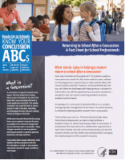
- School professionals play an important role in the health of all students. Recognizing the signs and symptoms of concussion is important, as is managing their return to school ...
- https://
- CDC's Concussion Training for Clinicians
-
Concussion Education Video Programs - Free and Easy
Parents, athletes, coaches and medical professionals have access to concussion education created...
-
New concussion guidelines for team physicians
INDIANAPOLIS – Team physicians who assess and treat athletes suspected of concussion have new ...

Surprising concussion myths and facts

Chris Hummel Clinical Associate Professor, Department of Exercise and Sport Sciences, Ithaca College


Current Events


quick links

iBaselineTM a division of ...

Knowing the signs and ...

Every school district and ...

Implement a concussion ...

Concussion – The Basics ...

Signs and symptoms of ...

What to do if a concussion is ...

Most concussions heal within ...
What to do if a concussion is ...

...

School medical ...

American Academy of Neurology ...
Latest News
Concussions Occur...
...in Any Sport
REMOVE athlete from play
REFER to medical provider
REST no sports, no texting/TV
RETURN only with doctor's OK
Source: Children's Hospital Boston, Sports Concussion Clinic