


![]()
![]()
![]() Join our newsletter!--------------------------------------------------------------------------------------------------
Join our newsletter!--------------------------------------------------------------------------------------------------
Does CTE infect neuron to neuron?


- Created on Saturday, 30 June 2012 09:56
- Last Updated on 13.09.2012
- Published Date
NFL Hall of Famer "Iron Mike" Webster's life ended in 2002 when he suffered a heart attack at age 50. Four Super Bowl rings, nine Pro Bowls, and voted to the NFL's all-time team in 2000, the driven, ultra-competitive Steelers' center drifted into a life of drug use, homelessness, and enormous suffering after retiring in 1988. In 2005, his son Garrett told ESPN that the only way his father could get to sleep was to taser himself into unconsciousness. Former teammates and friends offered assistance over the years but it wasn't enough.
After Webster's death, forensic neuropathologist Dr. Bennet Omalu was determined to find neurological reasons for his cognitive decline. Seventeen years in the NFL at a time when safety precautions and concussion awareness were both minimal had resulted in thousands of impacts to his head. Though his brain looked normal at autopsy, Omalu pressed on until he found the abnormalities that we now call Chronic Traumatic Encephalopathy, or CTE.
CTE and Alzheimer's
CTE and Alzheimer's are both “neurodegenerative diseases.” That is, they are brain diseases that result in the progressive destruction of brain cells, eventually leading to cognitive, behavioral, and other impairments. Both Alzheimer’s and CTE can only be accurately diagnosed postmortem through a neuropathological examination of the brain and their progression has remained elusive. But clues about how Alzheimer's spreads from neuron to neuron are now emerging. Whether the same process of progression applies to CTE is currently unclear.
Photo: Alzheimer's infects from neuron to neuron
The inexorable spread of Alzheimer’s disease through the brain leaves dead neurons and forgotten thoughts in its wake. Researchers at Linköping University in Sweden are the first to show how toxic proteins are transferred from neuron to neuron.
Through experiments on stained neurons, the research team – under the leadership of Martin Hallbeck, associate professor of Pathology – has been able to depict the process of neurons being invaded by diseased proteins that are then passed on to nearby cells.
“The spread of Alzheimer’s, which can be studied in the brains of diseased patients, always follows the same pattern. But until now how and why this happens has not been understood,” says Hallbeck, who along with his research group has now published their results in The Journal of Neuroscience.
The illness starts in the entorhinal cortex – a part of the cerebral cortex, and then spreads to the hippocampus. Both of these areas are important for memory. Gradually, pathological changes take place in more and more areas of the brain, while the patient becomes even sicker.
Two proteins have been identified in connection with Alzheimer’s: beta amyloid and tau. Normally tau is found in the axons – the outgrowths that connect between neurons – where it has a stabilising function, while beta amyloid seems to have a role in the synapses where the neurons transfer signal substances to each other. But in Alzheimer’s patients, something happens with these proteins; autopsies reveal abnormal accumulations of both.
Why they become abnormal is still unknown, but what is known is that it’s not the large accumulations, or plaques, that damage the neurons. Instead, smaller groups of beta amyloid – called oligomeres – seem to be the toxic form that gradually destroy the neurons and shrink the brain.
“We wanted to investigate whether these oligomeres can spread from neuron to neuron, something many researchers tried earlier but didn’t succeed,” Hallbeck says.
The study was inaugurated with an experiment on neuron cultures, where researchers injected oligomeres stained with a phosphorescent red substance called TMR using a very thin needle. The next day the neighbouring, connected neurons were also red, which showed that the oligomeres had spread.
To test whether a sick neuron can “infect” others, they conducted a round of experiments with mature human neurons stained green and mixed with others that were red after having taken up stained oligomeres. After a day, approximately half of the green cells had been in contact with a few of the red ones. After two more days, the axons had lost their shape and organelles in the cell nucleus had started to leak.
“Gradually more and more of the green cells became sick. Those that hadn’t taken up the oligomeres, on the other hand, weren’t affected,” Hallbeck says.
The study is a breakthrough in understanding Alzheimer’s and itshttps://ss. If a way of stopping the transfer can be found, it could lead to a more effective inhibitor against the disease.
https://Article: Spreading of neurodegenerative pathology via neuron-to-neuron transmission of beta-amyloid by Sangeeta Nath, Lotta Agholme, Firoz Kurudenkandy, Björn Granseth, Jan Marcusson and Martin Hallbeck. The Journal of Neuroscience, 27 June 2012.
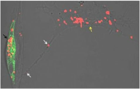
Photo: Alzheimer's infects from neuron to neuron: Two nerve cells, each about 10 micrometers large, are visible as shadows in this picture. From the beginning only the right one (yellow arrow) contained the toxic, red stained, oligomeric beta-amyloid. When these sick cells make contacts with the healthy, green labeled cells (black arrow), toxic beta-amyloid will spread through the neuronal projections (white arrow). Subsequently, also the green cell will become sick. Credit: Martin Hallbeck
Source: The above story is reprinted from materials provided by Linköping University.
Questions/comments? contact Jean Rickerson at This email address is being protected from spambots. You need JavaScript enabled to view it.
Brain Health

Children and young adults scanned multiple times by computed tomography (CT), a commonly used diagnostic tool, have a small increased risk of leukemia and brain tumors in the decade following their ...
read more...-
Why concussions affect people differently
Bronx, NY — Patients vary widely in their response to concussion, but scientists haven’t unde...
-
Teens miss recovery clues after concussion
PITTSBURGH — When recovering from concussion, young athletes rely too much on how they f...
-
Discovering the roots of migraine
Common questions encountered during the post-concussion exam are often migraine-related. Do you...


Neuroscience

NFL Hall of Famer "Iron Mike" Webster's life ended in 2002 when he suffered a heart attack at age 50. Four Super Bowl rings, nine Pro Bowls, and voted to the NFL's all-time team in 2000, the driven, ...
read more...-
Progesterone seems to protect neurons after injury
It is not yet known why girls suffer concussions at a higher rate than boys. The most prevalent...
-
911 signal relay sends help to brain injury
Like emergency workers rushing to a disaster scene, cells called microglia spe...
-
GPS for the brain; the "connectome"
Athens, Ga.- University of Georgia researchers have developed a map of the human brain that shows...
Resources

- School professionals play an important role in the health of all students. Recognizing the signs and symptoms of concussion is important, as is managing their return to school post-injury.
- Some ...
- https://
- CDC's Concussion Training for Clinicians
-
Concussion Education Video Programs - ...
Parents, athletes, coaches and medical professionals have access to concussion education created...
-
New concussion guidelines for team ...
INDIANAPOLIS – Team physicians who assess and treat athletes suspected of concussion have new ...

The risk of concussions in young football players

Marjorie Albohm President, National Athletic Trainers' Association


Current Events


quick links

iBaselineTM a division of ...

Knowing the signs and ...

Every school district and ...

Implement a concussion ...

Concussion – The Basics ...

Signs and symptoms of ...

What to do if a concussion is ...

Most concussions heal within ...
What to do if a concussion is ...

...

School medical ...

American Academy of Neurology ...
Latest News
Concussions Occur...
...in Any Sport
REMOVE athlete from play
REFER to medical provider
REST no sports, no texting/TV
RETURN only with doctor's OK
Source: Children's Hospital Boston, Sports Concussion Clinic